Complex Evolution of Human Dental Development
Citation
Smith, T.M.*, Tafforeau, P.*, Le Cabec, A., Bonnin, A., Houssaye, A., Pouech, J., Moggi-Cecchi, J., Manthi, F., Ward, C., Makaremi, M., Menter, C.G. (2015) Dental ontogeny in Pliocene and early Pleistocene hominins. PLOS ONE 10(2): e0118118. * These authors contributed equally to this work.
Link: http://dx.plos.org/10.1371/journal.pone.0118118
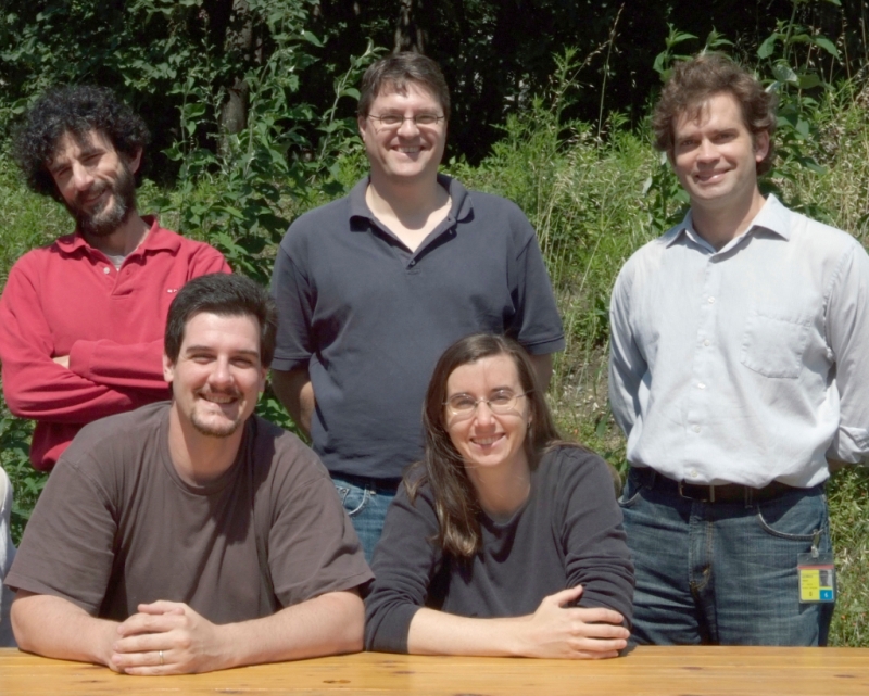
Scientists have been debating whether early human ancestors (hominins) followed human-like or ape-like rates of growth and development since the description of the enigmatic Australopithecus africanus Taung child in 1925. Fossil teeth are important for resolving this issue as dental microstructure preserves permanent records of birth, daily growth, developmental stress, and age at death in juveniles. Until recently, accessing these permanent microscopic features required physically cutting teeth to produce cross sections for light microscopy, which severely limits available samples. Over the past decade, Beamline Scientist Paul Tafforeau at the European Synchrotron Radiation Facility in Grenoble, France, and Harvard University Associate Professor Tanya Smith have worked to virtually resolve microscopic growth in fossil teeth (see Video 1 and Video 2). These studies have helped to document the earliest evidence for a modern human pattern of growth and development in a 160,000 year-old fossil Homo sapiens individual, and have demonstrated developmental differences between modern humans and their “evolutionary cousins” the Neanderthals. A new study by Smith, Tafforeau and colleagues (Figure 1) in the journal PLOS ONE details how synchrotron X-ray imaging was recently applied to more than 20 fossil hominins from East and South Africa in order to conduct the largest study of tooth development and age at death in early hominins.
Figure 1. Several members of the collaborative team in Grenoble, France. Top row left to right: Jacopo Moggi-Cecchi (Università di Firenze), Colin Menter (University of Johannesburg), Charlie Lockwood (formerly of University College London). Bottom row left to right: Paul Tafforeau (European Synchrotron Radiation Facilility) and Tanya Smith (Harvard University). This paper is dedicated to the memory of Charlie Lockwood (1970-2008), who passed away a month after this photo was taken.
Image credit: Chantal Argoud.
The Study in Brief
Over the course of this six-year study, biological rhythms in teeth (dental microstructure: Figure 2) were quantified with synchrotron X-ray imaging in a diverse sample of juvenile fossil hominins from the Pliocene and early Pleistocene (~1.5-4.2 million years ago). This allowed precise calculation of the duration of tooth formation and age at death in four hominin species: Australopithecus anamensis, Australopithecus africanus, Paranthropus robustus, and early Homo sp. The results reveal greater developmental variation than was previously appreciated. It is clear that all early hominins were not developmental alike; the enigmatic taxon Paranthropus robustus shows unique development (short crown formation times, late tooth initiation ages, and possibly later molar eruption ages) when compared to other early hominins. An additional finding is that South African early Homo teeth have surprisingly thick tooth enamel and rapid enamel secretion rates, unlike the condition in contemporaneous Asian Homo erectus, which underscores the need for additional recovery and study of these poorly understood South African fossils.
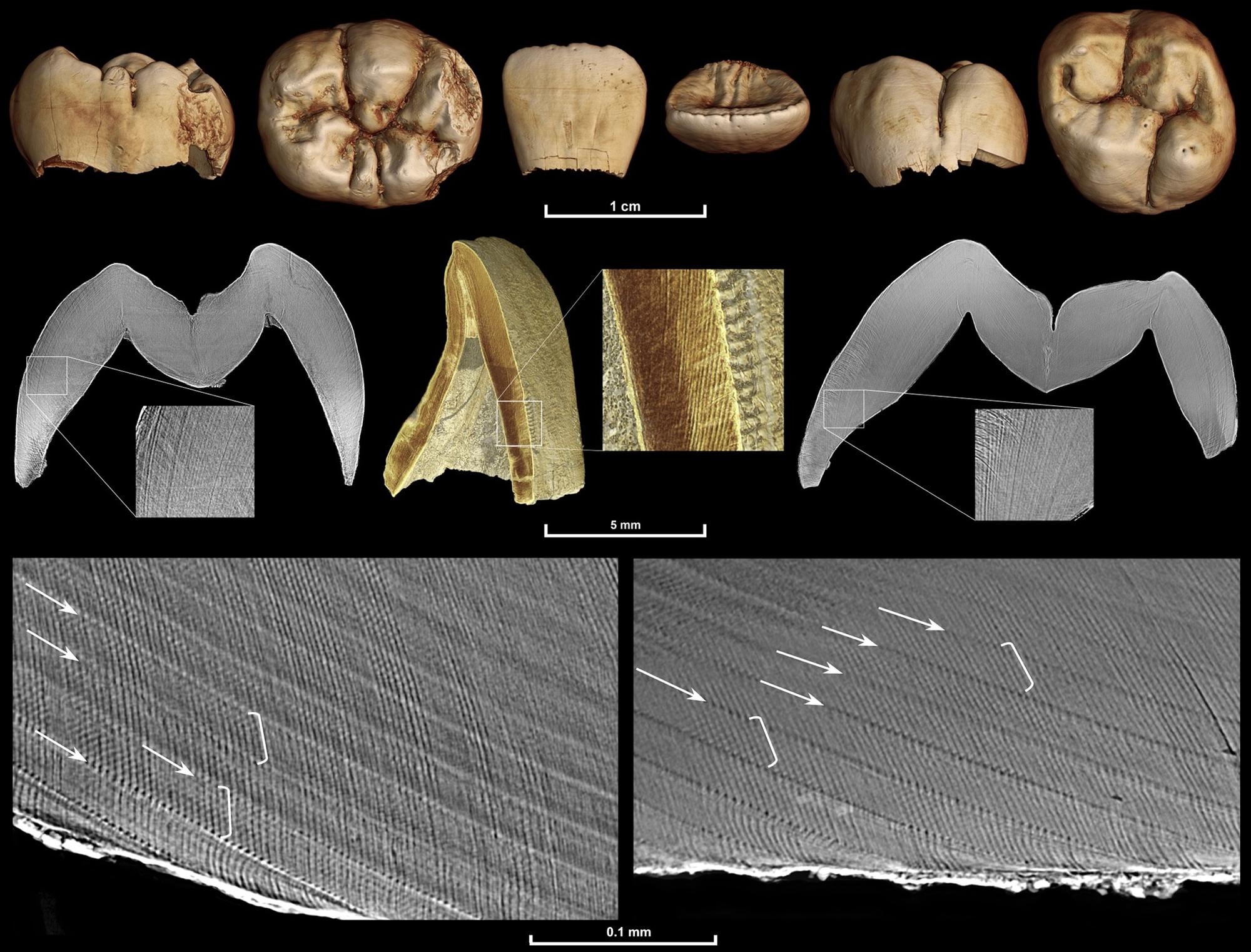
Figure 2. Multiscale synchrotron imaging of a fossil Homo juvenile (DNH 67/70/71).
Images are from scans performed with the following voxel sizes: 20 micron (upper row), 5 micron (middle row), and 0.7 micron (lower row). An identical internal developmental defect pattern confirmed that the two molars, which were found isolated but in close proximity, came from the same individual. They also both show an identical long-period line periodicity of 8 days, as 8 light and dark bands (daily cross-striations illustrated in white brackets) can be seen between successive long-period lines (Retzius lines illustrated by white arrows). It was not possible to determine the age death for this individual due to postmortem loss of the incisor cervix and dentine from all teeth.
Image credit: Smith et al. (2015) PLOS ONE.
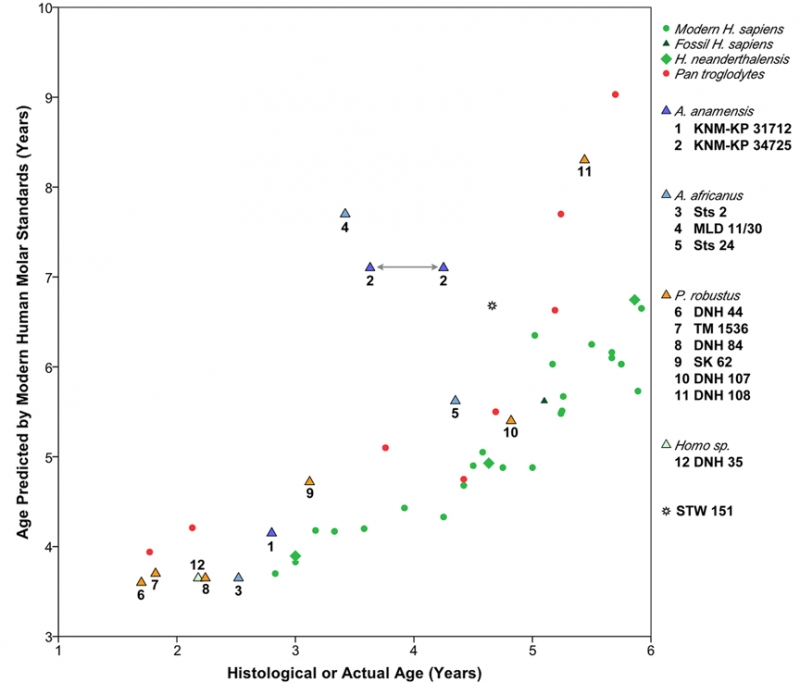
One of the most powerful outcomes of counting tiny growth lines in a juvenile’s dentition is the precise estimation of age at death, which has been proven to be highly accurate in living humans and non-human primates. This is possible due to the preservation of the “neonatal line,” a tiny stress line formed at birth that allows developmental time to be converted to chronological age. The current study reexamined several fossils that had been previously studied, as well as several newly discovered specimens, and found that several published age at death estimates are inaccurate. New chronological ages for two well-known fossils (SK 62 and StW 151) are several months younger than previous histological estimates (Figure 3), while the Australopithecus africanus individual Sts 24 is more than one year older than earlier reports suggest. Importantly, these ages were determined from developmental information quantified directly from the specimens themselves and supplemented with species-specific information when necessary. This facilitates more accurate age estimates and allows for independent developmental comparisons with living humans and chimpanzees. The results demonstrate that while the timing of tooth mineralization in most early hominins is more similar to chimpanzees than to living humans, several fossils appear to show even more rapid dental development than either living group (Figure 4).
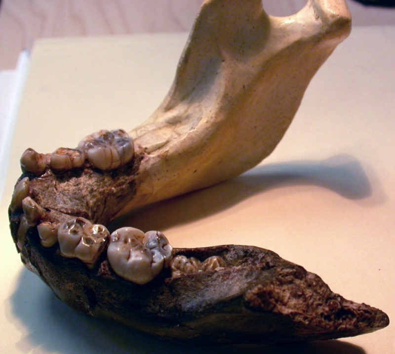
Figure 3. The 3.1 year-old Paranthropus robustus SK 62 child’s mandible, which is several months younger than a previous analysis that reported it died at 3.35-3.48 years. Note that the lower left first molar is still deeply embedded in the bone, suggesting that tooth emergence into the mouth would not have occurred for at least another few months. This specimen reveals a remarkably young age of central incisor eruption compared to living humans or chimpanzees, which do not erupt their incisors for several more years. The right side of the mandible consists of a plaster reconstruction that was added after the fossil was discovered in South Africa.
Image credit: Tanya Smith.
Significance
This study is the most comprehensive assessment of tooth growth, molar eruption, and age at death in australopiths and early Homo. A major strength of this approach includes precise assessments of internal developmental features, which have been poorly understood due to the historic need for physical sectioning to access this information. Non-destructive synchrotron investigations have greatly increased access to fossil specimens, allowing more comprehensive study of complete dentitions of extremely rare species. When compared with living humans and African apes, differences in Pliocene and early Pleistocene hominin incremental development and tooth calcification timing prohibit simplistic “human-like” or “ape-like” characterizations. For example, chimpanzee developmental standards predict that the 3.1 year old Paranthropus robustus individual SK 62 would have been 3.6 years of age when it died, while modern human standards suggest an age of 4.8 years old at death. In short, radiographic developmental standards from living humans and chimpanzees, or traditional analyses based on external features only, do not yield consistent or accurate ages at death for Pliocene or early Pleistocene hominins, and ontogenetic assessments of other juveniles such as the Taung child and Dikika infant should be reconsidered. These findings underscore the unique development of our own species, Homo sapiens, which mineralizes and erupts teeth more slowly than any other hominin, leading to our characteristically prolonged childhood.
Figure 4. Ages predicted from living human molar calcification standards (left, Y-axis) compared to known- or histologically-derived ages from this study (bottom, X-axis). Early hominins, which are represented by triangular symbols, fall closer to the chimpanzee sample (red circles) than the living human sample (green circles). Early hominins are more developmentally advanced than living humans, which show a long slow period of dental development.
Image credit: Smith et al. (2015) PLOS ONE.
Funding and Curating Institutions
This study was funded by the US National Science Foundation, the European Synchrotron Radiation Facility, the Radcliffe Institute for Advance Study, the Wenner-Gren Hunt Fellowship, Harvard University, the National Research Foundation of South Africa, the Italian Ministry of Foreign Affairs (Direzione Generale per la Promozione e la Cooperazione Culturale, ufficio V, Archaeology), and the Italian Embassy and Italian Cultural Institute in Pretoria. We are also grateful for the support of the DITSONG Museums of South Africa, National Museums of Kenya, South African Heritage Resources Agency, and the University of the Witwatersrand, which allowed these fossils to be imaged in Grenoble, France.
